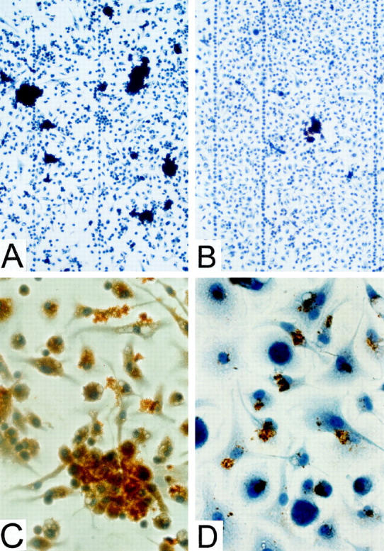
Figure 5.

Immunochemical detection of SAA/AA in cultures of adherent peritoneal cells after incubation with rSAA2 +/− AEF. A: Cells cultured with rSAA2 and AEF for 48 hours; relatively high density of immunostained (dark brown) areas seen under low magnification, ×100. B: Cells cultured with rSAA2 for 48 hours in the absence of AEF; relatively low density of immunostained areas seen under low magnification, ×100. C: Cells cultured with rSAA2 and AEF for 48 hours; magnification, ×400. All immunostaining is in contact with cells. D: Cells cultured with rSAA2 and AEF for 2 hours; magnification, ×600.
