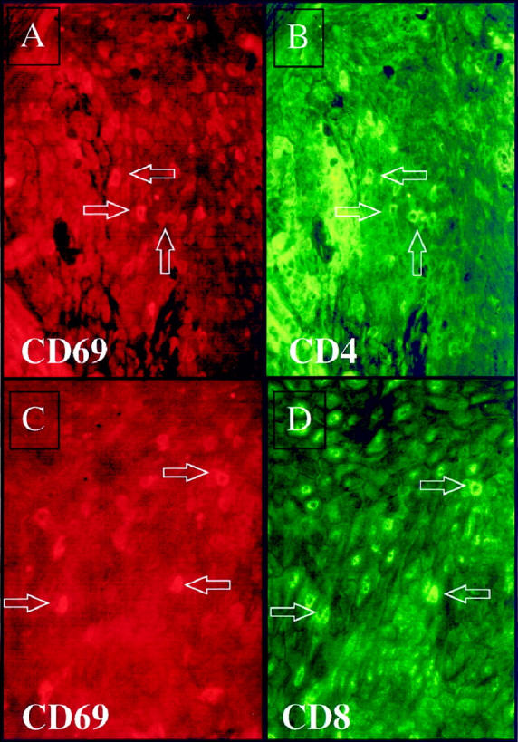
Figure 7.

Twenty-one days after injection of a CD4+ T cell line into PN skin, scattered intraepidermal immunocytes express rhodamine-stained CD69 (A and C). FITC-labeled antibody staining revealed that both CD4+ T cells (B), as well as CD8+ T cells (D) were CD69-positive. Arrows indicate double-positive cells.
