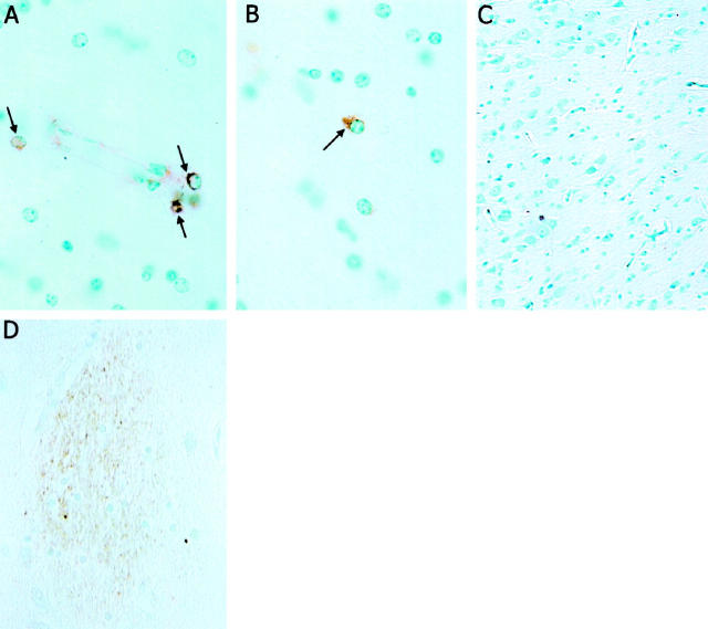
Figure 1.
Presence of Tat immunoreactivity in neurons in the brains of SHIV-infected macaques and HIV encephalitis patients. Formalin-fixed paraffin sections from the hippocampal formation of a SHIV-infected macaque with encephalitis (A and B), an uninfected macaque (C), and a patient with HIV encephalitis (D) were immunostained with Tat antibody (see Methods). Several Tat-immunoreactive cells are present in perivascular cells (A, arrows) and in white matter (B, arrow) of the SHIV-infected macaque, whereas no Tat-immunoreactive cells are present in the uninfected macaque (C). Tat immunoreactivity is prominent in microglial nodules in the section from the AIDS dementia patient (D). Similar results were obtained in analyses of sections from three HIV encephalitis patients, three SHIV-infected macaques, two human control patients, and two uninfected monkeys.

