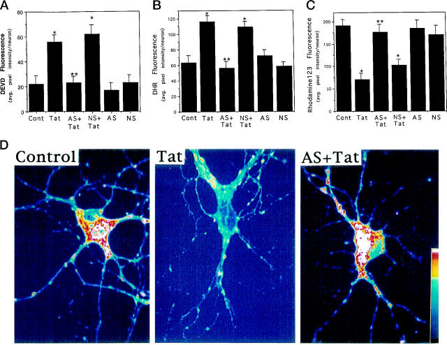
Figure 4.
Evidence that Par-4 induction mediates Tat-induced caspase activation, oxidative stress, and mitochondrial dysfunction in cultured hippocampal neurons. (A-C): Cultures were pretreated for 2 hours with 20 μmol/L Par-4 antisense (AS) or non-sense (NS) oligonucleotides and were then exposed for 6 hours to vehicle or 200 nmol/L Tat. Levels of DEVD fluorescence (A, a measure of levels of activated caspase-3), DHR fluorescence (B, a measure of levels of oxidative stress), and rhodamine 123 fluorescence (C, a measure of mitochondrial function) were quantified. Values are the mean and SE of determinations made in 4–6 cultures (15–25 neurons analyzed in each culture). *P < 0.01 compared to control value. **P < 0.01 compared to value for cultures exposed to Tat alone. Analysis of variance with Scheffé’s post hoc tests. D: Confocal laser scanning microscope images of rhodamine 123 fluorescence in hippocampal neurons from an untreated control culture, a culture exposed to 200 nmol/L Tat for 6 hours, and a culture preincubated for 2 hours with 20 μmol/L Par-4 antisense (AS) and then exposed to 200 nmol/L Tat for 6 hours. Note that Tat caused a decrease in the level of rhodamine 123 fluorescence, and that Par-4 antisense largely prevented the effect of Tat.

