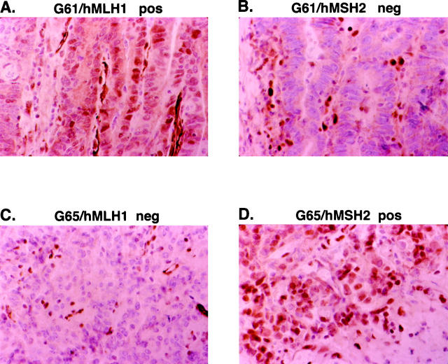
Figure 2.
Representative examples of loss of hMSH2 or hMLH1 expression in two gastric carcinomas (G61 and G65) with high-level instability (MSI-H). The neoplastic cells in the tumor of case G61 show a loss of hMSH2 (B) expression but normal hMLH1 (A) protein expression in the neoplastic cells. The neoplastic cells in the tumor of case G65 display a loss of hMLH1 (C) but normal hMSH2 (D) protein expression in the neoplastic cells. Lymphocytes that show strong nuclear staining for hMSH2 and hMLH1 serve as positive internal controls (B and C).

