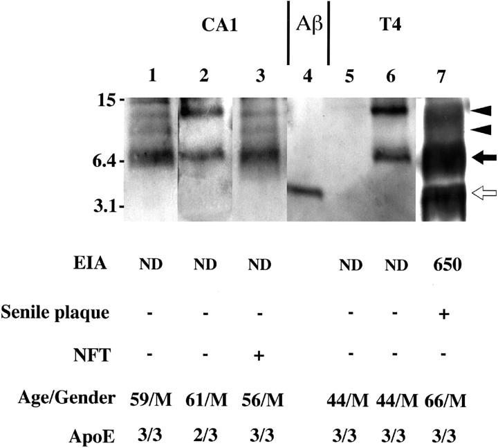
Figure 2.
Representative Western blots of the insoluble fraction of hippocampus CA1 (lanes 1 to 3) and T4 (lanes 5 to 7) homogenates. The formic acid extract (representing Aβ in the insoluble fraction) was subjected to Western blotting with BC05. Immunocytochemical scores regarding NFTs and SPs, insoluble Aβ42 levels quantitated by BNT77-based EIA (pmol/g wet weight), age/gender, and ApoE genotypes are also given. BNT77-quantitated Aβ corresponds to Aβ monomers on the Western blot. Lane 4, 50 pg of synthetic Aβ 1-42. ND, not detected (below the detection limit). The numbers in the left indicate molecular weights of marker proteins in kilodaltons. Open arrow , closed arrow, and arrowheads in the right indicate Aβ monomer, Aβ dimers, and putative Aβ oligomers, respectively.

