
Figure 2.
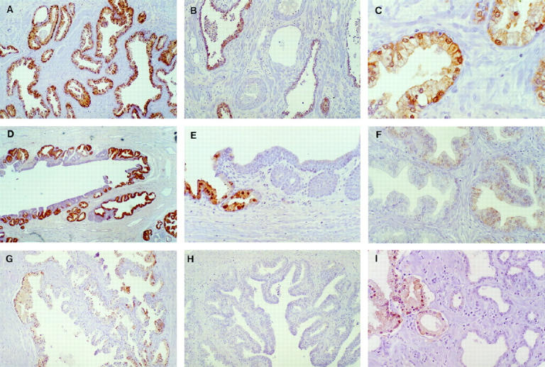
15-LOX-2 immunostaining in benign prostate. A: Uniform strong staining of glandular epithelium (original magnification, ×200). B: Absent immunostaining in vein and artery (middle), fibromuscular stroma, and admixed chronic inflammatory cells. Strong staining of glands in right and left of field (×100). C: Higher magnification showing cytoplasmic granular and focal nuclear staining of apical (secretory) epithelial cells (×400). D: Staining of secretory cells in large prostatic duct (×100). E: Higher magnification showing positive staining in columnar secretory cells (left) and lack of staining in transitional epithelium (right) of prostatic duct (×200). F: Non-uniform and reduced intensity staining in hyperplastic glands in transition zone (×200). G: Focal staining of glands in central zone (region around ejaculatory ducts toward base of gland; ×100). H: Complete absence of staining of ejaculatory duct (lumen toward lower right; ×100). I: Absent staining in atrophic glands (right). Compare with positive immunostaining of more normal-appearing glands, with ample cytoplasm, in left of photomicrograph (×200). Paraffin immunoperoxidase staining using rabbit polyclonal anti-15-LOX-2, 1:2500.