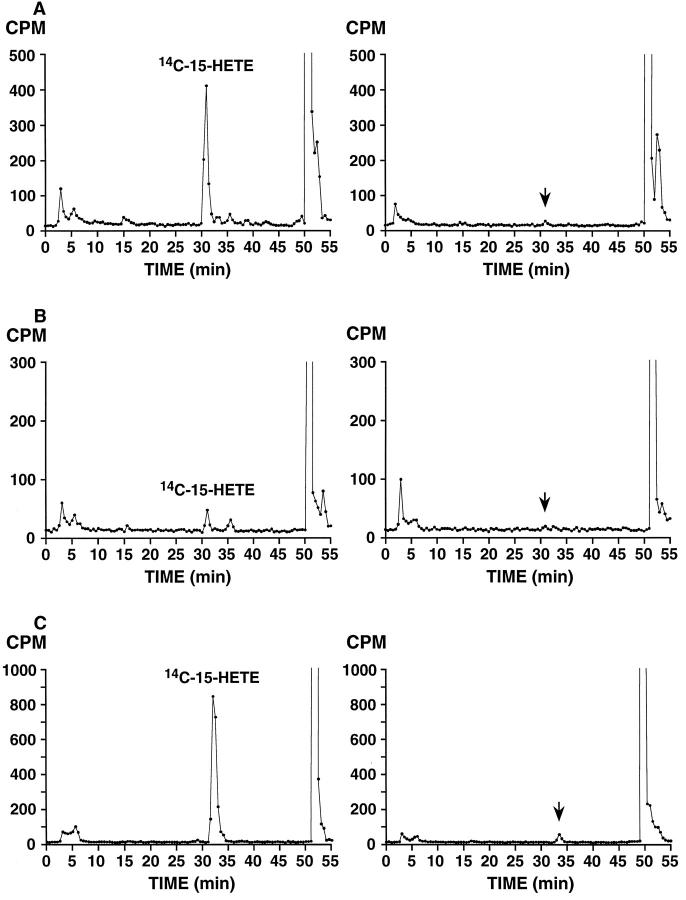
Figure 3.
15-HETE formation by benign (left) and malignant (right) prostate tissue from three different radical prostatectomy specimens. A: Benign and Gleason grade 3 + 4 = 7, with extra-capsular extension; B: Benign and Gleason grade 4 + 3 = 7, confined to prostate; C: Benign and Gleason grade 3 + 2 = 5, confined to prostate (with incubated tumor tissue from Gleason pattern 3 tumor in peripheral zone). Incubation with [14C]AA and extraction were performed as described in Materials and Methods. Product analysis was by reverse-phase HPLC using a Beckman Ultrasphere 5-μm ODS column (25 × 0.46 cm) with a solvent of methanol/water/glacial acetic acid (75:25:0.01, by volume) at a flow rate of 1.01 ml/minute switched to 100% methanol at 40 minutes; 0.5-minute fractions were collected and subjected to scintillation counting. [14C]15-HETE peaks are indicated in benign incubations and arrows corresponding to substantially reduced or undetected [14C]15-HETE in tumor incubations based on retention time of unlabeled HETE standards co-injected with 14C samples.

