
Figure 2.
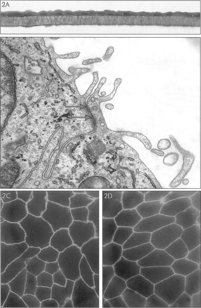
Characterization of RPE monolayers cultured on transwell filter for 3 weeks. A: Light microscopy of RPE cell monolayer (donor B), stained with hematoxylin-eosin. B: Electron microscopy of the apical side of the RPE cell monolayer. Note the presence of apical microvilli and between the cells a structure (indicated by arrows) suggesting the presence of a tight junction. C and D: Immunofluorescence microscopy of RPE cell monolayers of donor B (low TER: 35 Ohm·cm2) (C) and donor D (high TER: 100 Ohm·cm2) (D) stained for tight junction-associated protein ZO-1. Note staining for ZO-1 at the intercellular junctions.