
Figure 3.
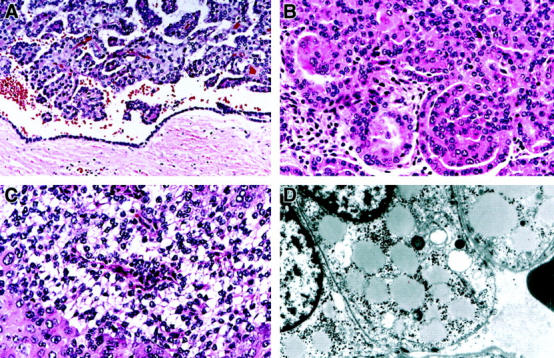
Histopathology of representative PRC and an ultrastructure of clear cells in hereditary PRC patients 5946 with V1110I mutation and 4599 with H1112R mutation. A: Fibrous pseudocapsule surrounds a 4 cm tumor from patient 5946; a retraction artifact gives an impression of a “cyst wall lined by a layer of tumor cells” (H&E, ×200). B: A tumor from patient 4599 shows a “metanephric adenoma-like” architecture and contains cells with low grade basophilic nuclei and amphophilic and eosinophilic cytoplasm (H&E, ×400). C: A tumor from patient 5946 demonstrates an area composed of sheets of cells with basophilic Fuhrman nuclear grade 1 nuclei and clear cytoplasm and adjacent cells with Fuhrman nuclear grade 3 nuclei and eosinophilic cytoplasm. Thin fibrovascular papillary cores are seen in cross-section in the area of clear cells (H&E, ×400). D: Electron micrograph of clear cells from a hereditary PRC patient 5946 shows prominent intracytoplasmic lipid droplets and glycogen (magnification, ×6600).