Table 2.
Summary of Reconstructed Vascular Lesions
| Case no. and block | Diagnosis | Total length of reconstruction (μm) | Branching pattern (axial* or dichotomous†) | Sequence and spacing of vascular changes after branch point (N, P, C, or D × distance in μm) |
|---|---|---|---|---|
| 1a | PPH | 445 | Dichotomous | Branch 1: C× 230, P× 200 |
| Branch 2: P× ? | ||||
| 1b | PPH | 245 | Dichotomous | Branch 1: N× 35, C× 105, P× 100 |
| Branch 2: medial hypertrophy× ? | ||||
| 1c | PPH | 170 | Dichotomous | Branch 1: N× 35, C× 65, P× 70 |
| Branch 2: medial hypertrophy× ? | ||||
| 1d | PPH | 250 | Dichotomous | Branch 1: N× 40, C× 55, P× 80 |
| Branch 2: N× 30, P× 145 | ||||
| 2 | PPH | 260 | Dichotomous | Branch 1: N× 25, P× 120, D× 80 |
| Branch 2: N× 35, P× 110, D× 80 | ||||
| 3 | Cirrhosis | 410 | Axial | P× 400, D× ? |
| 4 | AIDS | 540 | Dichotomous | Branch 1: N× 200, P× 70 |
| Branch 2: N× 55, P× 200 | ||||
| 5a | Scleroderma | 340 | Axial | N× 10, C× 50, P× 300 |
| 5b | Scleroderma | 340 | Dichotomous | Branch 1: P× 200 |
| Branch 2: N× 35, C× 70, P× 100 | ||||
| 6 | Normal | 145 | Dichotomous | Branch 1: N |
| Branch 2: N |
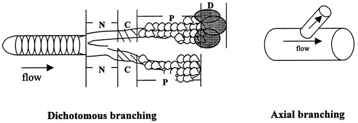
*Axial = A medium-sized axial pulmonary artery gives rise to a branching, smaller-sized artery.
†Dichotomous = A normal-appearing vessel bifurcates into two equally sized branches.
N = Normal pulmonary artery.
C = Concentric-obliterative lesion.
P = Plexiform lesion.
D = Dilatation lesion.
? = Vessel disappears in distal sections.

