
Figure 1.
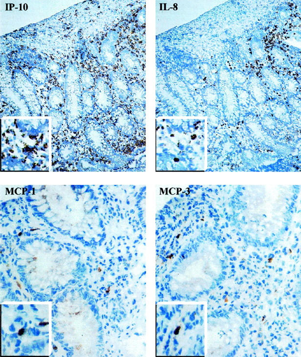
Micrograph of serial sections from a representative UC colonic mucosa stained for IP-10, IL-8, MCP-1, and MCP-3. Higher magnification of chemokine-positive cells are shown in the insets. Original magnifications, ×20 (IP-10 and IL-8) and ×40 (MCP-1 and MCP-3).