
Figure 2.
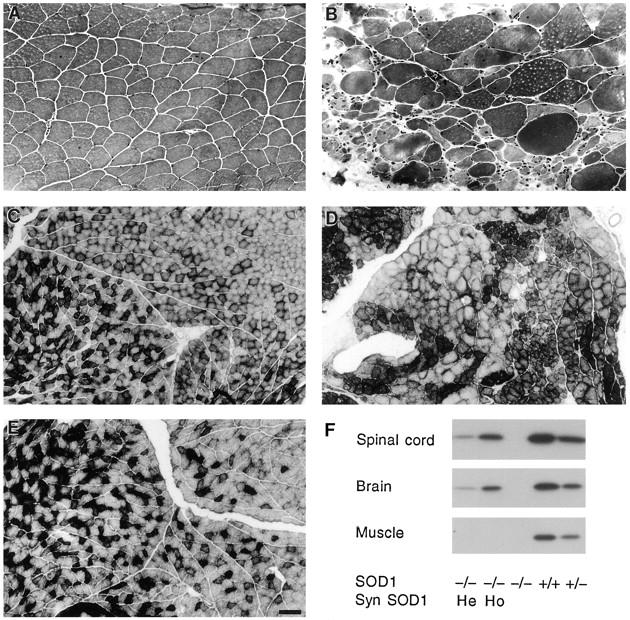
Hindlimb muscles showing denervation in SOD1−/− mice and Cu/Zn SOD protein levels. Medial gastrocnemius muscle from a 6-month-old SOD1+/+ mouse (A) showing normal muscle fibers, and from a SOD1−/− mouse (B) showing marked atrophy of muscle fibers consistent with denervation. Some muscle fibers in B are hypertrophied. Hematoxylin and eosin stain. Central gastrocnemius muscle from a 12-month-old SOD1+/+ mouse (C) showing the normal interspersion of type IIA and IIB fibers. Same muscle from a 12-month-old SOD1−/− mouse (D) showing fiber type grouping, consistent with chronic denervation and reinnervation and from a 12-month-old SOD1−/−, hemizygous synapsin SOD1 transgenic mouse (E) showing a normal pattern of muscle fibers. Succinic acid dehydrogenase histochemistry. Bars, 50 μm (A and B) and 125 μm (C-E). F: Cu/Zn SOD protein levels in brain, spinal cord, and hindlimb muscle in SOD1+/+, heterozygous (SOD1+/−), and SOD1−/− mice. SOD1−/− mice were either wild-type, hemizygous (He), or homozygous (Ho) for the synapsin (Syn) SOD1 transgene, which directed Cu/Zn SOD expression to brain and spinal cord but not to muscle.