
Figure 2.
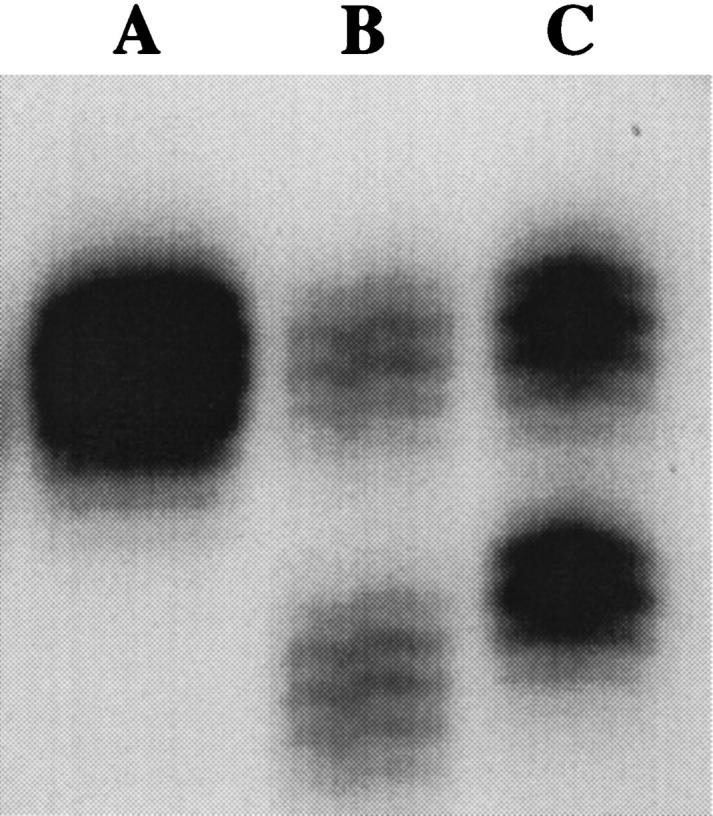
Results from the amplification of the BAT-25 locus from normal male lymphocyte DNA (Lane A), colorectal tumor DNA exhibiting MSI (Lane B) , and lymphocyte DNA from one representative healthy African-American (Lane C). The banding pattern in Lane A depicts the allelic pattern of 25 thymine repeats previously described for BAT-25 in normal MSS DNA. Lane B (MSI-positive tumor) illustrates the characteristic banding pattern of MSI seen at BAT-25 with one band corresponding to alleles with shortened mononucleotide repeats (lower band) compared to a second band demonstrating an allele of wild-type repeat length (upper band). Lane C depicts the banding pattern seen in blood DNA from one representative African-American with one allele within the previously described range of size (upper band) and one polymorphic 17-repeat allele (lower band).