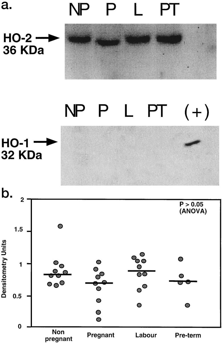
Figure 2.

a: Western blot analysis for HO-2 (upper panel) and HO-1 (lower panel) in human nonpregnant myometrium (NP), pregnant term nonlaboring myometrium (P), pregnant term laboring myometrium (L), and preterm laboring myometrium (PT). Each lane was loaded with 25 μg of membrane protein. A band of 36 kd was detected in the membrane fraction (M) of all of the samples with the HO-2 antibody. No HO-2 was detectable in the cytosolic fraction (C) of any of the samples. A positive control (+) for HO-1 (recombinant rat HO-1) confirmed the HO-1 antibody’s specificity. HO-1 was undetectable in all of the samples. b: Scanning densitometric analysis of autoradiographs obtained from non-pregnant (n = 10), pregnant term nonlaboring (n = 10), term laboring (n = 10), and preterm nonlaboring (n = 5) myometrium. Data are shown as a scattergram and bars show median values.
