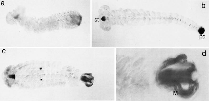
Figure 3.
RNA expression of Tc-fkh. Embryos were stained by whole-mount in situ hybridization. a–c represent progressively older stages, and d is an enlargement of the posterior end of an embryo that is at the same stage as the embryo in c. st, stomodaeum; pd, proctodaeum; M, Malpighian tubules. Note that the dots seen in the middle of the embryo in c are unspecifically staining cells in the pleuropodia on segment A1.

