
Figure 3.
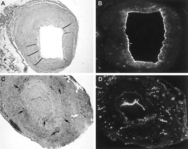
Giant cell formation, fragmentation of the internal elastic lamina, and neoangiogenesis in GCA. Temporal arteries from GCA patients with and without marked neoangiogenesis were compared. Hematoxylin/eosin (A, C) and von Willebrand’s factor staining (B, D) of two representative specimens are shown. Original magnification, ×100. B and C: In the temporal artery with vasa vasorum confined to the adventitia, multinucleated giant cells were absent and the internal elastic lamina (IEL), was well maintained. C and D: In the temporal artery with numerous microvessels in the media and the hyperplastic intima, a garland of multinucleated giant cells (arrows) was arranged along the disrupted IEL.