
Figure 3.
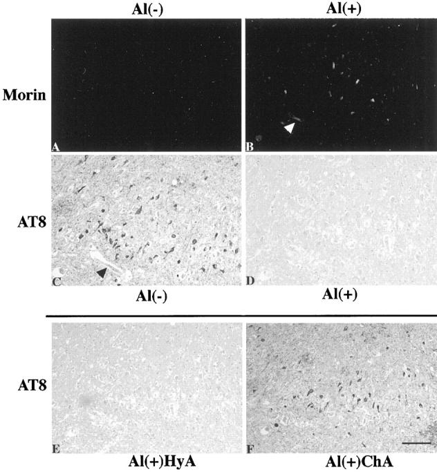
The Al accumulation with association to NFTs and the Al-induced immunoreactive abolishment of the phosphorylated epitope in PHFτ of the NFD. Closely adjacent sections of the AD hippocampus were pretreated by different sets of procedures before Morin staining (A and B) or immunostaining with AT8 (C-F). A and B: The sections were subjected to prior Al removal by the chelating autoclave method to differentiate binding of exogenous Al. They were then incubated with control buffer without Al (A) or with buffer containing 10 mmol/L AlCl3 pH 6.5 (B). C-F: The sections were initially treated by the hydrated autoclave method as the routine antigen retrieval. They were then incubated with control buffer without Al (C) or with buffer containing 10 mmol/L AlCl3, pH 6.5 (D-F), followed by incubation in control buffer without DFO (C and D), by the hydrated autoclave method (HyA) (E) or by the chelating autoclave method with 30 mmol/L DFO, pH 4.7 (ChA) (F). Incubation with AlCl3 produced Al accumulation with association to NFTs (B) and abolished the immunolabeling of the NFD revealed by AT8 (D). This immunoreactive abolishment was retrieved by the chelating autoclave method (F), but not by the hydrated autoclave method (E). Arrowheads in B and C indicates the same blood vessel. All panels are at the same magnification. Scale bar, 50 μm.