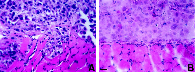
Figure 1.
Photomicrographs of tumors of the prostate carcinoma cell lines PC-3N (A) and DU-145 (B) on the surface of diaphragms of SCID mice. SCID mice (n = 4) were injected intraperitoneally with 5 × 10 5 cells, sacrificed 5 weeks after injection, and the diaphragms fixed and processed in paraffin. DU145 tumors have penetrated the basement membrane, and PC-3N have penetrated through the murine straited muscle. Five-micron sections were cut and deparaffinized for hematoxylin and eosin staining. Scale, 60 μm.

