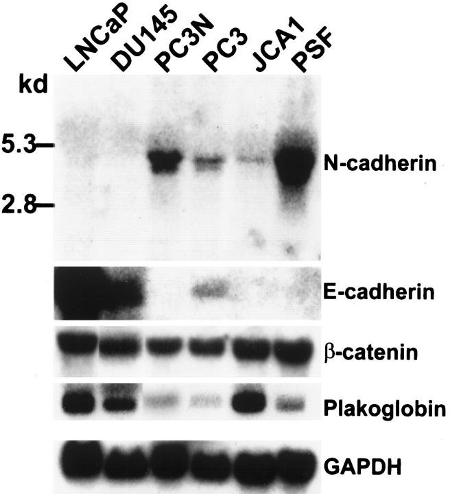
Figure 4.
Northern blot analysis of E- and N-cadherins and plakoglobin in prostate carcinoma cell lines and prostate stromal fibroblasts. Twenty micrograms per lane of total RNA from each cell line was blotted on a nylon membrane. A 300-bp EcoR I fragment to N-cadherin was used as a probe for N-cadherin detection. A 1.7-bp fragment to mouse E-cadherin was used as a probe for E-cadherin expression. Full-length plakoglobin and β-catenin cDNA were used to detect plakoglobin and β-catenin expression. A 1.2-kb GAPDH cDNA fragment was used as normalization standard. The hybridized membranes were exposed to X-ray films for 1 day (E-cadherin, β-catenin, plakoglobin, GAPDH) and 2 days (N-cadherin). This is a representative result of two independent experiments.

