
Figure 4.
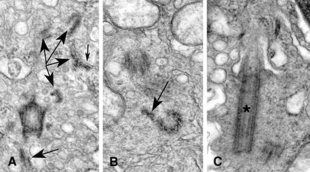
Abnormal centriole structure in breast tumors. A: Subdistal appendages are seen in this oblique section through a centriole. Numerous microtubule complexes (large arrows) are seen in various planes of section throughout the cytoplasm near the centriole. As is seen in cross section of the complexes, the individual microtubules share a portion of the wall of the neighbor microtubules (small arrow). B: The open-ring configuration of this centriole is shown in cross section. Two of the nine triplet microtubule complexes are splayed away from the centriole barrel (arrow). C: This centriole bearing a primary cilium (*) is nearly twice as long as normal centrioles. Original magnifications, ×54,500 (A), ×59,625 (B), ×47,700 (C).