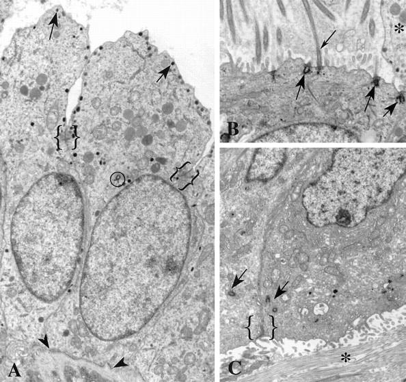
Figure 5.

Positional centrosomal anomalies in breast tumors. A: Secretory granules (arrows) are present at the apical membrane of these cells displaying apocrine metaplasia. Junctional complexes (brackets) mark the transition from lateral to apical membrane domains. Apocrine beaks extend into the lumen of the duct. Notice the centriole (circled) near the apical end of the nucleus. These cells have apical/basal polarity and rest on a basement membrane (arrowheads). B: Extra centrioles in this cell are inserted at the apical plasma membrane where they function as basal bodies (large arrows) for cilia (small arrow). Microvilli and cilia project into the lumen. The beak of an adjacent apocrine cell (*) is visible. The ciliated cell does not protrude into the lumen, as does the apocrine cell; but like its apocrine neighbor, it has apical/basal polarity and rests on a basement membrane (not visible in this figure). C: The two centrosomes (arrows) seen in adjacent cells are located near the junctional complex between these polarized cells (bracket). However the apical membrane domain with microvilli faces collagen (*) of the stromal tissue rather than the lumen of a duct. This invasive group of cells has ramified through the breast stroma and is not subtended by a basement membrane. The polarity of these cells is inverted, with the basal domains abutting the basal domains of other cells and the apical domains facing the stroma rather than a lumen. Original magnifications, ×8150 (A), ×10,000 (B), ×7900 (C).
