Figure 1.
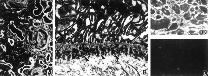
A and B: GLA renal biopsy tissue. A: Immunofluorescence micrograph demonstrates immunostaining for κ light chain in glomerular and tubular basement membranes. B: Electron micrograph of a tubule shows clustered granular electron-dense deposits (arrows) in basement membrane. C and D: CHO myocardial tissue. C: Immunofluorescence micrograph reveals perimyocytic deposits staining for κ. D: No staining for λ light chain. Magnification: A, ×200; B, ×10,000; C and D, ×300.

