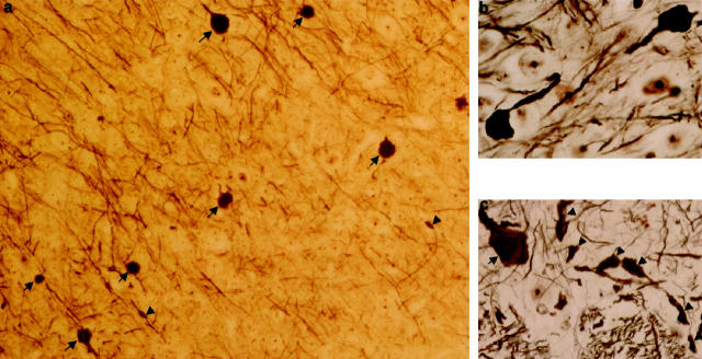
Figure 3.
Silver staining of brain and spinal cord of homozygous htau40 transgenic mice at the age of 2.5 months. a: Multiple dilated axons or axonal spheroids (arrows) and some irregular dystrophic axons (arrowheads) in cortex (×360). b: Higher magnification of two dilated axons in thalamus. Note that the dilations approach the size of neuronal cell bodies (×920). c: Aspect of spinal cord gray matter (anterior horn) with a grossly dilated axon (arrow) and several irregularly thickened dystrophic axons (arrowheads) (×390).

