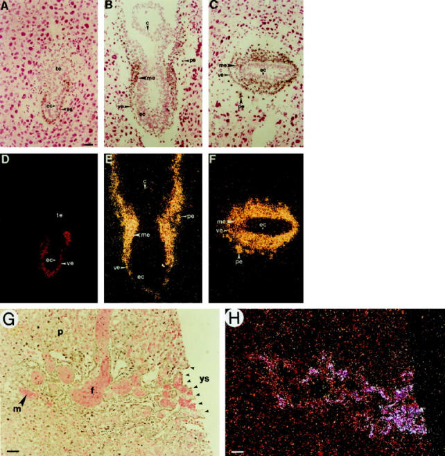
Figure 1.

In situ hybridization of GATA-4 mRNA in mouse embryos. Corresponding bright-field (A–C, G) and dark-field (D–F, H) views are shown. A and C: Longitudinal section through a 6-day p.c. embryo. B and E: Longitudinal section through a 7-day p.c. embryo. C and F: Transverse section through a 7-day p.c. embryo. G and H: Section through the chorionic plate of a 14-day p.c. embryo. Note that GATA-4 mRNA is abundantly expressed in the visceral and parietal endoderm of the postimplantation embryo. Later, some expression is seen in the nascent embryonic mesoderm, but not in ectoderm or trophectoderm. In the late gestation embryo, GATA-4 mRNA is expressed by yolk sac endoderm cells within the “Crypts of Duval.” c, chorion; ec, ectoderm; f, fetal blood sinus; m, maternal blood sinus; me, mesoderm (nascent); p, placental cells (decidua); pe, parietal endoderm; te, trophectoderm; ve, visceral endoderm; ys, yolk sac. Original magnification: A–H, ×200. Scale bar = 20 μm.
