
Figure 1.
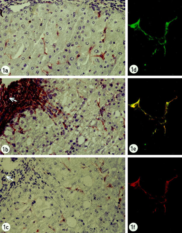
a: Liver biopsy of patient with chronic biliary disease showing synaptophysin reactivity in perisinusoidal cells with stellate projections. Original magnification, ×250. b: Liver biopsy of patient with chronic hepatitis C showing α-smooth muscle actin-reactivity in lobular perisinusoidal stellate cells, in septal myofibroblasts (left upper side of figure) and in smooth vessel cells of an arterial wall (arrow) Original magnification, ×250. c: Liver biopsy of patient with chronic hepatitis C showing synaptophysin-immunoreactivity in lobular perisinusoidal cells with stellate projections. In the fibrotic septum (upper left side of figure), no reactivity is present. Arterial wall, arrow. Original magnification, ×250. d-f: Liver biopsy of patient with chronic hepatitis C: double staining for synaptophysin and α-smooth muscle antigen, detected by the confocal laser scanning microscope: Lobular stellate cell showing reactivity for synaptophysin (green labeling, d), α-smooth muscle actin (red labeling, f) and colocalization for synaptophysin and α-smooth muscle actin (yellow double-labeling, e). Original magnification, ×1000.