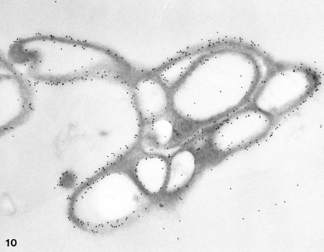
Figure 10.
Cyrosection of another normal cell prepared after exposure to hypertonic conditions. The frozen thin section was stained for GPIb in the same manner as the cells in Figures 8 and 9 ▶ ▶ . Gold particles indicating the presence of GPIb are dispersed evenly on the exposed surface (S) and in the OCS. Differences in gold particle frequency on outer and inner membranes are not apparent. Original magnification, ×42,000.

