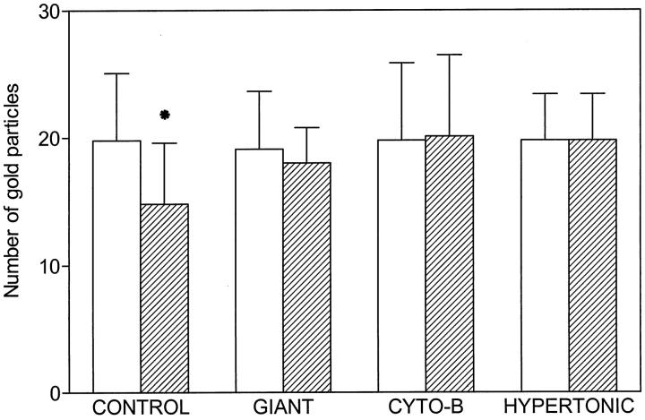
Figure 11.
Comparison of the number of gold particles on external (white bars) and internal membranes (dashed bars) of control, giant, cytochalasin B-treated, and hypertonically treated platelets. Densities of labeling are expressed in particles/μm. Statistical differences were observed for labeling on external versus internal membranes in control platelets (*P < 0.05). Differences were not observed under conditions where the open canalicular system was naturally or artificially dilated.

