
Figure 4.
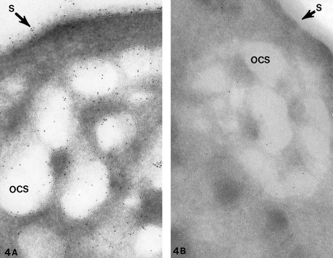
Cryosections of giant platelets from (A) a patient with Epstein’s syndrome (ES) and (B) a patient with Bernard Soulier syndrome (BSS). The cells were stained with the anti-glycocalicin antibody and protein A gold in the same manner as the cell in Figure 3 ▶ for the presence of GPIb. Gold particles indicating sites of GPIb cover the exposed surface (S) and membranes lining channels of the dilated OCS. There is no difference in the frequency of gold beads detecting GPIb on external or internal membranes of the giant ES cell. The specificity of the antiglycocalicin antibody for GPIb is indicated by the virtual absence of immunogold particles on and in the BSS platelet in B. Original magnifications, ×100,000 (A) and ×70,000 (B).