
Figure 6.
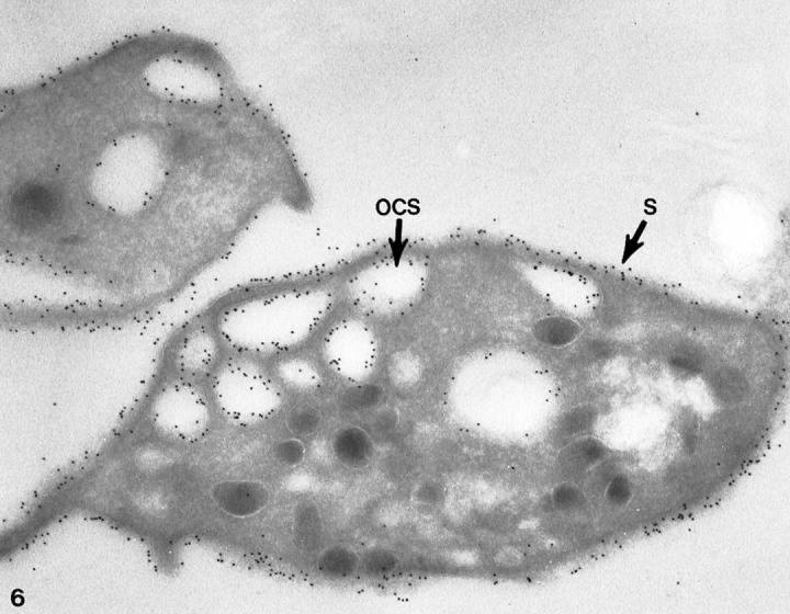
Cryosection of a normal platelet incubated with cytochalasin B stained for GPIb in the same manner as the cell in Figure 5 ▶ . Immunogold beads detecting sites of GPIb are prominent on membranes of the cell surface (S) and lining the OCS. The receptors appear to have the same density on internal and external membranes. Original magnification, ×42,000.