
Figure 7.
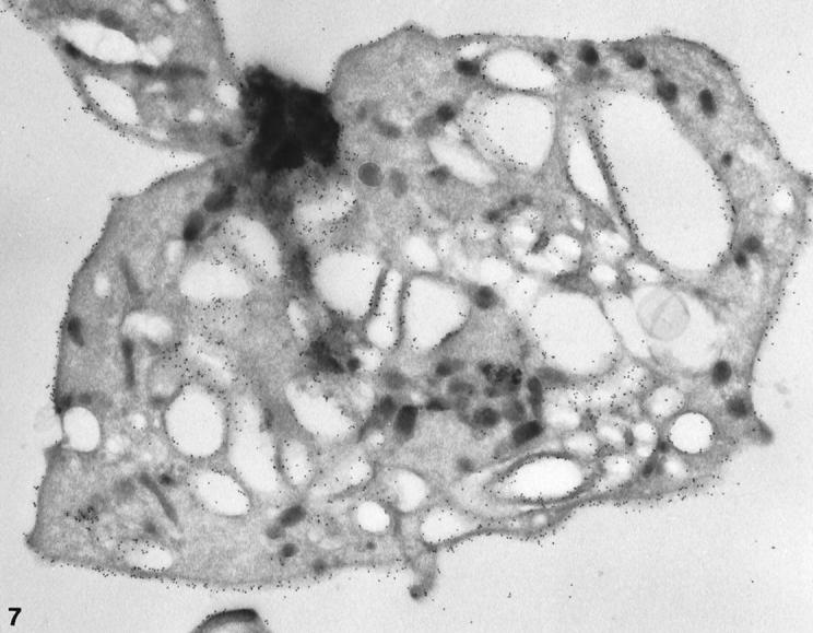
Cryosection of a larger normal platelet with a dilated OCS prepared after exposure to CB. The frozen thin section was stained with AP1, 6D1, and anti-mouse IgG coupled to 10 nm gold in the same manner as the cell in Figure 5 ▶ . Immunogold beads indicating sites of GPIb cover the external and internal membranes. The frequency of gold particles is essentially the same on the exposed surface (S) and membranes lining OCS channels. Original magnification, ×42,000.