Figure 5.
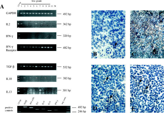
A: Semiquantitative RT-PCR analysis of cytokine expression in MALT-type lymphoma tissues. N, negative control. Th1-weighted cytokines were normally absent, despite weak expression of IL2 in cases 1, 6, and 8. In contrast, Th2/Th3-weighted cytokines (TGF-β1, IL10, and IL13) were present in low-grade lymphomas. Ionomycin-stimulated normal PBLs from healthy donors were taken as positive controls. Protein expression in immunohistochemistry paralleled mRNA expression. B: Only a few IL2-positive cells (arrow) (V, vessels) with a faint cytoplasmatic staining were found perivascularly and within the tumor tissue (case 1; PAP, cryostat section, ×400). C: Strong cytoplasmic staining for TGF-β1 in tumor cells as well as in small blood vessels (case 6; PAP, cryostat section, ×200). D and E: IL10 (case 6; PAP, cryostat section, ×250; D) and IL13 (case 7; PAP, cryostat section, ×200; E). Numerous positive brown staining cells (arrows) were found loosely distributed within the tumor tissues.

