
Figure 1.
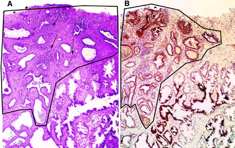
Proliferative inflammatory atrophy (PIA) of the prostate. A: Low-magnification view of a focus of PIA (outlined area) occurring adjacent to benign normal appearing glands (lower right). Arrows indicate collections of chronic inflammatory cells (predominantly lymphocytes). This lesion was classified as having marked chronic inflammation. H&E, ×40. B: Section adjacent to that shown in A, stained with anti-GSTP1 polyclonal antibody. Immunoperoxidase, ×40.