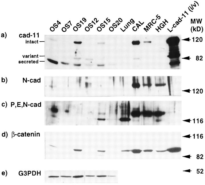
Figure 2.
Protein expression of cadherins and β-catenin in osteosarcoma. Lysates from six osteosarcoma samples were tested by Western blotting with an anti-cadherin-11 antibody (a), anti-N-cadherin antisera (b), an anti-P,E,N-cadherin antibody (c), an anti-β-catenin antibody (d), and an anti-G3PDH antibody (e). Each lane contains 40 μg of protein. Cell lysates from both the intact and variant forms of L cell transfectants (L-cad11 (i/v)) were loaded, and three forms of cadherin-11, the intact (120 kd), the variant (85 kd), and the secreted (80 kd) forms, were detected. Lysates from human calvarial cells (CAL), the human lung fibroblast cell line (MRC-5), and the human Giralidi heart cell line (HGH) were loaded as positive controls.

