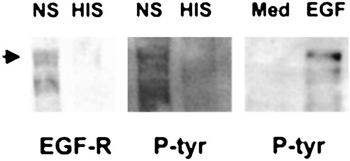
Figure 2.
Complement induces Grb2 association with EGF-R. Left and middle panels: GEC were incubated with antibody and normal serum (NS), or heat-inactivated serum (HIS) in controls, as in Figure 1 ▶ . Right panel: GEC were incubated with EGF (100 ng/ml, 60 minutes, 37°C). Cell lysates were incubated with agarose-conjugated GST-Grb2 fusion protein and subjected to SDS-PAGE and immunoblotting with antibodies to EGF-R (left panel) or to phosphotyrosine (P-tyr; middle and right panels). The arrow points to the 170-kd EGF-R. The lower band in the left panel probably represents an EGF-R degradation product.

