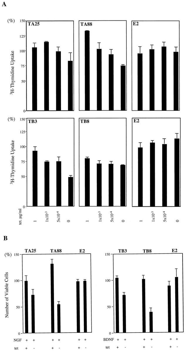
Figure 5.

Effects of NGF and BDNF on cell proliferation. A: NGF treatment of trkA transfected clones decreased [3H]thymidine uptake with high trkA expression (in the absence of tetracycline). The values are quoted as a percentage of the control (without NT treatment). The cells were cultured in RPMI with 10% FBS for 3 days at the different tetracycline concentrations, which ranged from 0 to 1 μg/ml, and were labeled with 1 μCi of [3H]thymidine for 20 hours. Radioactivity was measured on day 4 after NT treatment (100 ng/ml). Interestingly, TA88 showed increased [3H]thymidine uptake with low trkA at a tetracycline concentration of 1 μg/ml. B: Viable cell numbers are quoted as a percentage of the control (without NT treatment). The cells were treated with NGF or BDNF (100 ng/ml) for 5 days in RPMI with 10% FBS in the presence (1 μg/ml) and absence of tetracycline. tet +: 1 μg tetracycline/ml; tet −: 0 μg tetracycline/ml.
