
Figure 1.
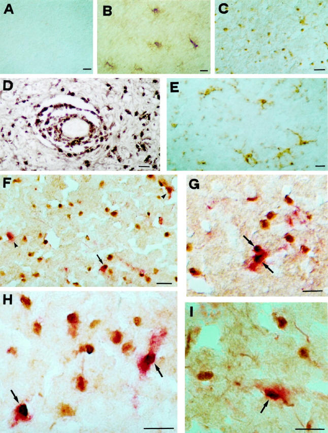
A to C: Immunoperoxidase staining for the activated NF-κB p65 subunit in normal CNS and MS lesions. A: No reactivity on glial cells is seen in white matter from control cases. B: Signal for NF-κB is visible in hypertrophic astrocytes in chronic silent MS lesions. C: At the edge of chronic active MS lesions, immunostaining for activated NF-κB shows nuclear reactivity on glial cells morphologically resembling oligodendrocytes or microglia. D: In addition to parenchymal cells, intense immunoperoxidase staining is detectable on perivascular, inflammatory elements. E: At variance with the pattern obtained for NF-κB, staining for its inhibitor, IκB, is detectable in the cytoplasm of microglia in the white matter surrounding MS plaques. F and H: Double-staining for activated p65 subunit of NF-κB (peroxidase, brown) and MBP (alkaline phosphatase, red) in chronic active MS lesions, reveals that a small proportion of NF-κB-positive nuclei present at the edge are surrounded by MBP-reactive cytoplasm (arrows), whereas most cells display no staining for MBP, probably representing microglial elements. About half of the oligodendrocyte population shows no NF-κB nuclear translocation (arrowheads). G and I: A pattern similar to NF-κB is detectable also for activated JNK at the edge of active MS lesions, where a proportion of MBP-positive oligodendrocytes (alkaline phosphatase, red; arrows) shows nuclear reactivity for JNK (peroxidase, brown). Scale bar, 30 μm.