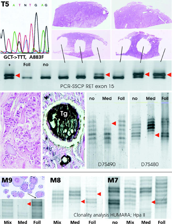
Figure 2.

Mutation analysis (top) and loss of heterozygosity analysis (middle) of microdissected medullary and follicular components from a mixed medullary-follicular carcinoma (T5). Note that in the medullary carcinoma portion the single-strand conformation analysis of RET exon 15 exhibits aberrant band patterns (red arrowheads) in a subset of the tumor (which is indicative of the presence of a somatic A883F missense mutation; upper left) but not in the follicular component (Foll) or nontumorous tissue (no). Note also that a loss of heterozygosity at the microsatellite loci D7S490 and D7S480 is only detectable in the medullary portion (Med) of the tumor (red arrowheads) and not in the microdissected thyroglobulin-positive (Tg) follicular part (Foll). Bottom, M9, M8, M7: Comparison of the X-chromosomal inactivation (clonality) pattern in the microdissected medullary carcinoma and follicular components of three mixed medullary-follicular thyroid carcinomas from informative female patients (for details see Materials and Methods). Note that all three tumors exhibit a monoclonal pattern in the medullary portion (Med), which is defined by the loss of one allele after HpaII digestion of DNA (red arrowheads), whereas a polyclonal pattern similar to that in normal tissue (no) or whole tumor extract (Mix) is encountered in the follicular component (Foll) of two tumors (M9 and M7), and a monoclonal pattern with inactivation of the opposite allele is present in the third tumor (M8).
