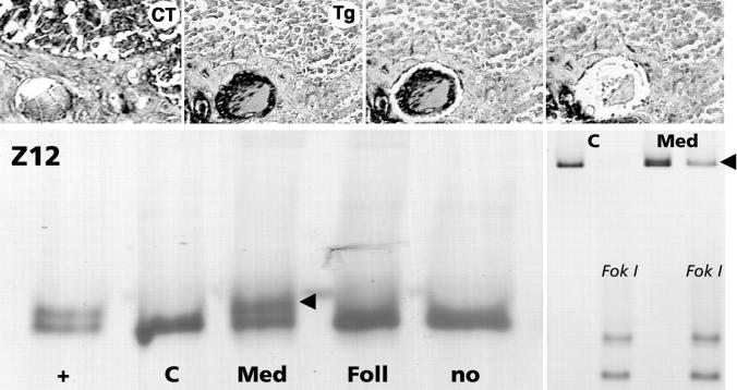
Figure 3.
Mutation analysis of RET exon 16 in the microdissected calcitonin-positive (CT) medullary and thyroglobulin-positive (Tg) follicular portions of a mixed medullary-follicular thyroid carcinoma (lymph node metastasis; Z12). Note that a heteroduplex formation (lower left, red arrowhead) is only detectable in the medullary (Med) but not in the follicular (Foll) tumor component. The presence of a somatic M918T point mutation was confirmed by FokI restriction analysis of PCR products (lower right, red arrowhead).

