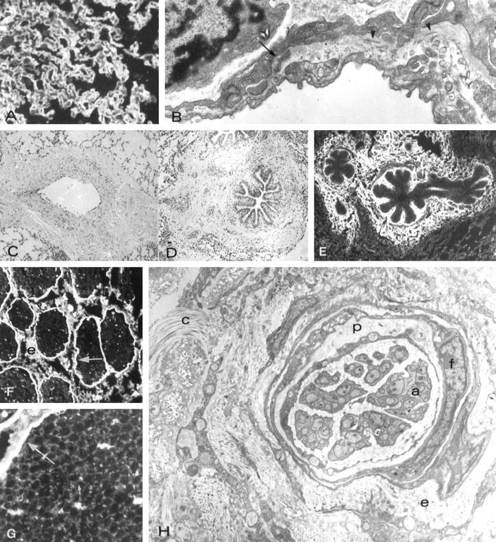
Figure 4.

Morphology and immunopathology in lungs of pigs Group III. (Pig N09) A: Thickened alveolar capillary walls stained by anti-laminin antibody. B: Electron micrograph of a thickened alveolar basement membrane containing a dense deposit (arrow) and collagen fibrils (arrowheads). C: Mural and adventitial sclerosis of a pulmonary vein. D: Peribronchial sclerosis. E: Type I collage in the bronchial lamina propria and in the peribronchial matrix. F: Binding of baboon anti-αGal to perineurium (arrow) and epinerium (e) uf unmyelinated nerve. G: Binding of baboon anti-αGal to the perineurium (large arrow) and endoneurium (small arrow). H: Electron micrograph showing the thickened and distorted perineurium (p) and sclerosis of the endoneurium (e), which contains increased amount of collagen (C). f, indicated fibroblast-liek perineurial cells; a, islands of unmyelinated axons. Original magnifications, A, C, D, E, ×300; B, ×20,000; F, ×600; G, ×1000; H, ×25,000.
