
Figure 5.
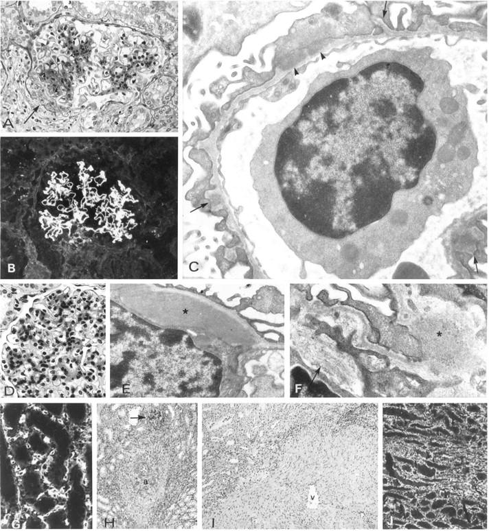
Morphology and immunopathology in kidneys of Group III pigs.
Lesions in the kidneys of pigs 11 (A–F) and 12 (G–I). A: Mesangial sclerosis and glomerulo-capsular adhesion (arrow). B: Deposits of baboon IgG in the glomerular capillary walls. C:: Electron micrograph showing glomerular basement membrane spikes (arrows), and subepithelial deposits of foreign material (arrowheads). D: Mesangial cell proliferation and sclerosis. E and F: Electron micrographs showing mesangial deposits (asterisks) and bundles of fibrils (arrows). G: Binding of baboon anti-αGal to fibroblasts in the inner stripe of the medulla, which strongly express αGal epitopes. H: Sclerosis around a medullary artery. The arrow indicates a glomerulus. I: Sclerosis around a medullary vein (v). J: Deposits in type I collagen in the medulla. A, B, D, G, ×600; C, E, F, 25.000; H, I, J ×200.