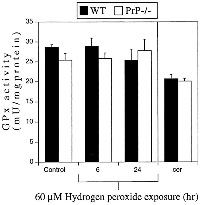
Figure 3.
GPx activity in WT and PrP−/− neurons. Lysates from CGN cultures or 2-week-old cerebellum (cer) from WT or PrP−/− mice were used to determine GPx enzyme activity. There was no significant difference in GPx activity between WT and PrP−/− neurons in control cultures or in cultures exposed to H2O2 (60 μmol/L) for 6 or 24 hours. No difference in GPx activity was observed between WT and PrP−/− cerebellum.

