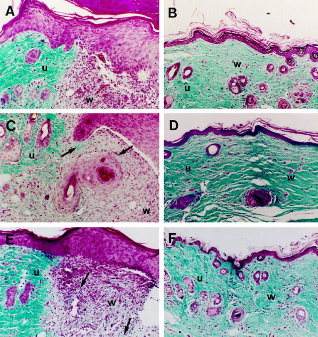
Figure 3.

Cutaneous wound healing in thrombomodulin-deficient mice. No differences in rate or extent of reepithelialization were observed between TM+/+ (A, B), TM+/− (C, D), or TMpro/− (E, F) mice. On day 7, foci of increased staining for collagen (arrows) were apparent in the wound matrices of TM+/− (C) and TMpro/− (E) mice compared to TM+/+ (A) mice. On day 30, dense collagen staining was observed in all three groups of mice, but collagen bundles tended to be thicker in TM+/− (D) and TMpro/− (F) mice than in TM+/+ mice (B). u, unwounded skin; w, wound matrix. Masson’s trichrome stain; magnification, ×150.
