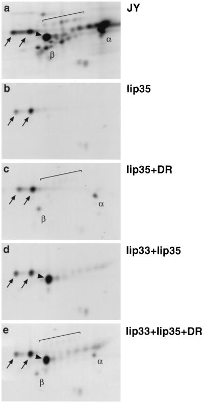
Figure 1.
Iip35 can exit the endoplasmic reticulum if associated with DR molecules or Iip33. Cells were labeled with [35S]-methionine for 2 hr, followed by a 2-hr incubation in nonradioactive medium before lysis and immunoprecipitation with mAb PIN.1.1. Samples from JY cells (a); or HeLa cells transfected with Iip35 (b); Iip35 and DR3 (c); Iip33 and Iip35 (d); or Iip33, Iip35, and DR3 (e) were analyzed by two-dimensional gel electrophoresis. Arrows indicate the core-glycosylated Iip35-spots, brackets the sialylated Iip35 spots. Core-glycosylated Iip33 is indicated by an arrowhead. α, DRα; β, DRβ. Acidic proteins are located to the right, basic proteins to the left.

