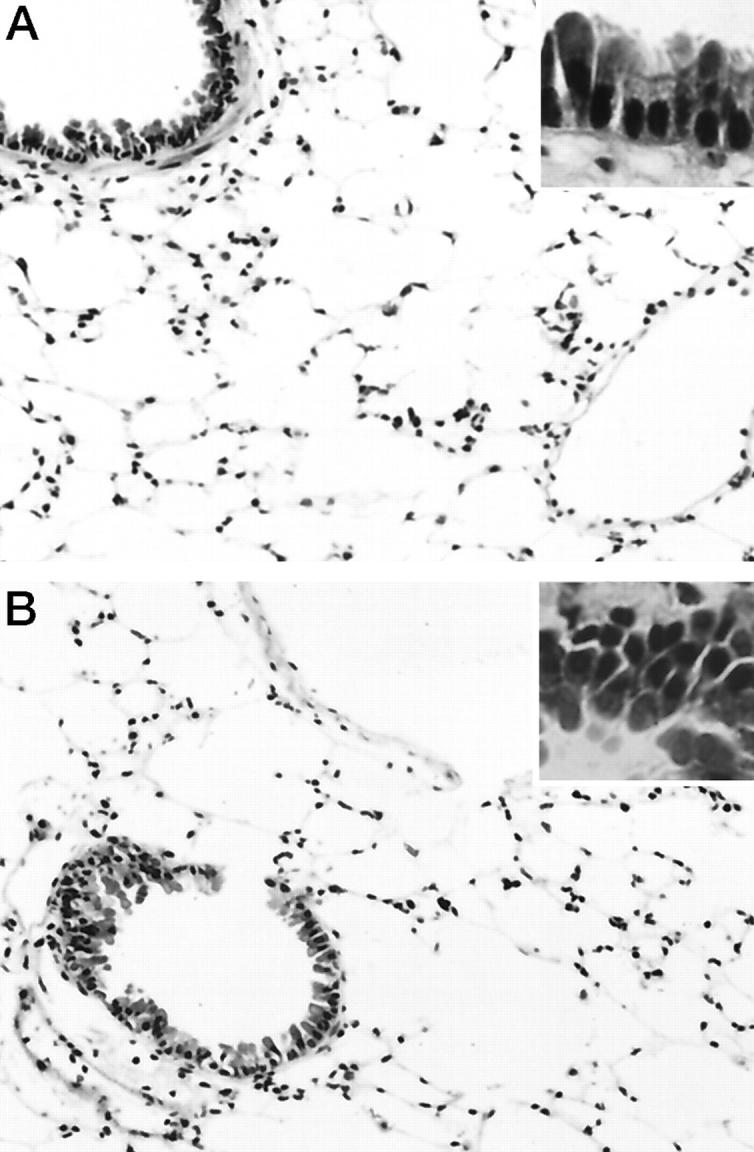
Figure 1.

Histological appearance of the lung from a wild-type (A) and p53 knockout (B) mouse at 3 days after the treatment. In both the wild-type and p53 knockout mice, a few inflammatory cell infiltrates composed of neutrophils and mononuclear cells were observed around bronchi and small blood vessels and within alveolar spaces. No cells exhibiting apoptotic characteristics were seen in either bronchi or alveoli of either group of mice (insets). Hematoxylin and eosin; original magnification, ×200 (insets, ×400).
