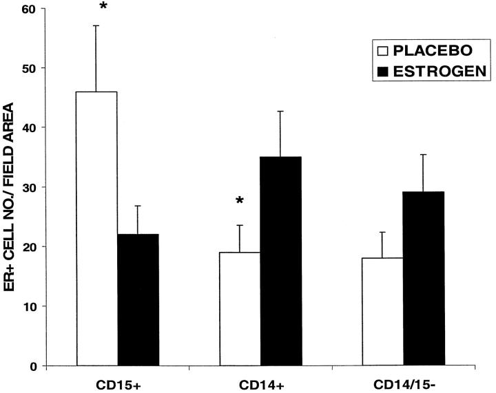
Figure 4.
Estrogen receptor localization in acute wounds. Quantification of estrogen receptor-positive (ER+) cells in specific subgroups at day 7 post-wounding showed a significant increase in ER+CD15+ (neutrophil) cells and decrease in ER+ CD14+ (monocyte) cells in the placebo versus estrogen-treated wounds. *P < 0.05, n = 5 per group. ER+ CD14− CD15− cells were considered to be fibroblasts on morphological grounds and were found to be increased (not significantly) in the estrogen-treated wounds. No gender differences were observed. The data represented graphically are that of the male groups.

