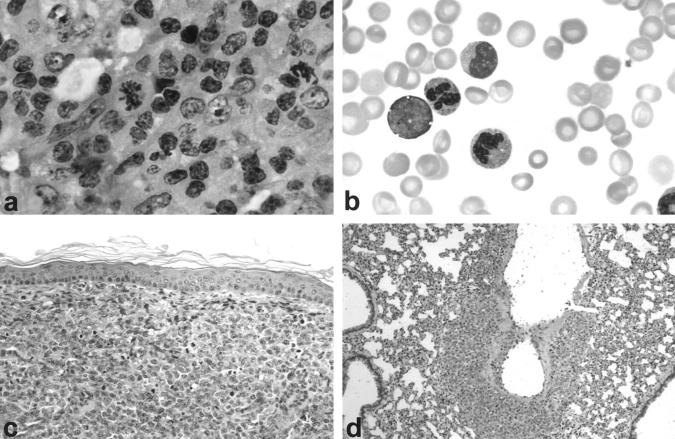
Figure 1.
a: Lymph node histology of ALCL patient. H&E, ×600. b: Circulating ALCL tumor cells in the peripheral blood. Giemsa stain, ×1000. c: Human ALCL tumor with anaplastic morphology infiltrating dermis of SCID/bg mouse. H&E stain, ×200. d: Tumor cells infiltrating walls of a pulmonary blood vessel. H&E stain, ×200.

