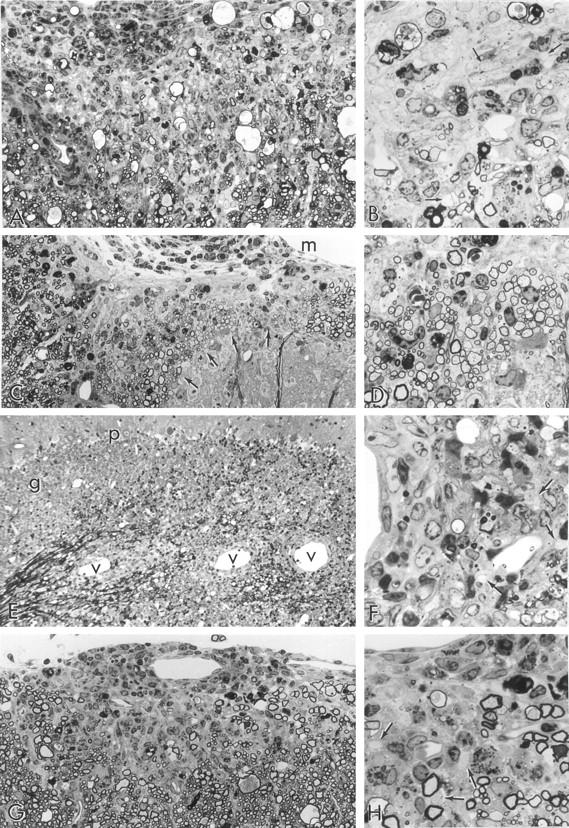
Figure 4.

Light micrographs taken from 1-μm epoxy sections stained with toluidine blue. A: Two-week-old recipient; 52 dpt; L7 spinal cord; clinical signs 10 days. A large area of anterior column shows chronic demyelination, glial scarring, infiltrating cells (mostly within the meningeal space, upper left), and large myelin ovoids. The latter are the residue of fibers that have undergone Wallerian degeneration. Original magnification, ×300. B: Detail from A showing demyelinated axons (arrows), background fibrous astrogliosis, residual macrophage activity and a few thinly myelinated (remyelinated) axons. This appearance is fairly typical of a long-standing lesion and suggests that changes in this CNS preceded clinical signs by several weeks. Original magnification, ×750. C: Two-week-old recipient; 52 dpt; L6 spinal cord; clinical signs 10 days. An area of white matter overlying a dorsal horn (lower right) displays collections of nerve fibers (arrows), with disproportionately thin myelin sheaths, an appearance typical of remyelination. Original magnification, ×300. D: Detail of an area of remyelination shown at one of the arrows in C. Note the large group of uniformly thinly myelinated (remyelinated) fibers. m = meningeal surface. Original magnification, ×750. E: Eight-week-old recipient; 10 dpt; cerebellum; clinical signs 3 days. Note lesions typical of acute EAE visible as perivascular cuffs of infiltrating cells around blood vessels (v) along the white matter of this cerebellar folium. Granule (g) and Purkinje (p) cells are also seen. Original magnification, ×300. F: Detail of a lesion from E showing demyelinated axons (arrows) in relationship to a perivascular cuff (left). Some apoptotic nuclei are apparent. Original magnification, ×750. G: Eight-week-old recipient; 52 dpt; cervical spinal cord; clinical signs 42 days, recent relapse (3 days earlier). An intensely infiltrated, small demyelinating lesion is seen in the subpial region of spinal cord, overlaid by a perivascular cuff around a vessel in the leptomeninges. Such an acute lesion occurring in this chronically afflicated animal correlates with the recent relapse. Original magnification, ×300. H: Detail from G showing demyelinated axons (arrows) within an edematous, nonfibrous astrogliotic parenchyma. Macrophages containing myelin debris are present. Original magnification, ×750.
