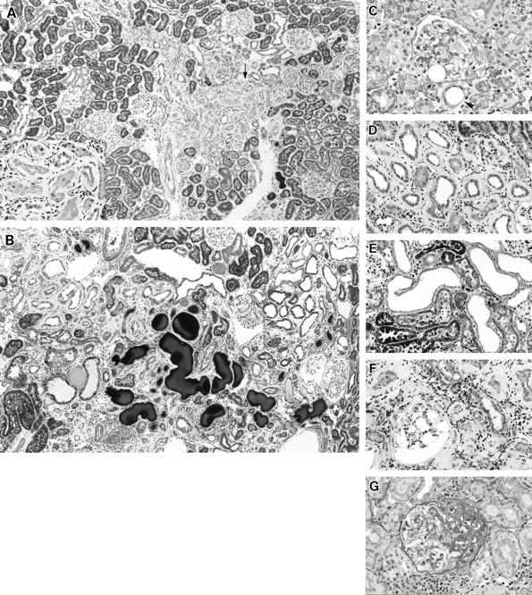
Figure 4.

Light micrographs of kidneys 4 to 12 weeks after microsphere injection in group 2 (0.8 mg of microspheres). A: Four weeks after microsphere injection, a zone of atrophic tubules with surrounding fibrosis is evident between islands of apparently intact proximal tubules (arrow). Periodic acid-Schiff (PAS) stain; original magnification, ×60. Inset: A higher magnified micrograph of the indicated area showed atrophic tubules surrounded by thickened basement membrane. Original magnification, ×130. B: Twelve weeks after microsphere injection, large dilated tubules with or without cast formation are seen scattered among atrophic tubules. Masson trichrome stain; original magnification, ×60. C: Four weeks after microsphere injection. Transparent round microspheres are observed in the interstitium (arrowheads) and at the vascular pole of a glomerulus (arrow). The glomerulus containing the microsphere appears essentially normal. PAS stain; original magnification, ×260. D: Eight weeks after microsphere injection, a cuff-like, thickened basement membrane is more prominent, and infiltration of round cells is evident around atrophic tubules. Trichrome stain; original magnification, ×130. E: Eight weeks after microsphere injection, large dilated tubules consist of short, flattened epithelial cells without a thickened basement membrane. Trichrome stain; original magnification, ×130. F: Twelve weeks after microsphere injection, the thickness of the tubular basement membrane is increased. A glomerulus shows huge empty vacuolar change in glomerular epithelial cells. Trichrome stain; original magnification, ×130. G: Twelve weeks after microsphere injection, a glomerulus shows segmental glomerular sclerosis with hyalinosis and adhesion to Bowman’s capsule. Trichrome stain; original magnification, ×220.
