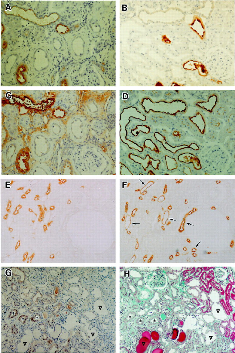
Figure 6.

Histochemical staining with anti-THP antibodies (A, B, and E), biotin-labeled PNA (C, D, and F) and anti-PCNA antibodies (G) on kidneys from animals in group 2. Sections in A, B, and E are cut serially to ones in C, D and F, respectively. A: Atrophic tubular cells showed no staining for THP, 8 weeks after microsphere injection. Original magnification, ×90. B: Some dilated tubules stained for THP, but not all at 12 weeks. Original magnification, ×90. C: Atrophic tubular cells were not stained by PNA. Section cut serially to (A). Original magnification, ×90. D: Almost all dilated tubular cells were stained by PNA. Section cut serially to B. Original magnification, ×90. E and F: Serial sections of the deep cortex in the kidney from a rat before microsphere injection (normal control) were stained with antibodies to THP (E) or biotin-labeled PNA (F) PNA clearly stained many distal tubules (arrows) as well as THP-positive ones. Brush borders of proximal tubules were only faintly stained. Original magnification, ×90. G and H: Twelve weeks after microsphere injection, serial sections were stained with antibodies to PCNA (G) or Masson-Trichrome stain (H). PCNA-positive cells are evident only in tubules with a cuff-like, thickened basement membrane (*). Large dilated tubules (▿) did not include positive cells. Original magnification, ×60.
